Figure 1.
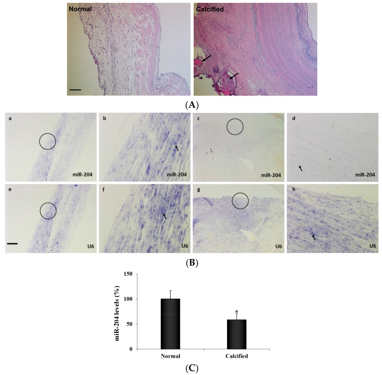
MiR-204 levels are lower in diseased human aortic valves. (A) Hematoxylin and eosin stain images (10× objective) of normal and calcified human aortic valve leaflets show that calcified aortic valve leaflets loss tri-layered morphology. Calcification nodules (arrows) are present in moderately calcified aortic valve leaflets. Scale bar = 100 µm. (B) Normal and calcified human aortic valve tissue sections were incubated with a full length DIG-labeled LNA probe to miR-204 (a–d) and a DIG-labeled LNA probe specific for the non-coding small nuclear RNA U6 as internal control (e–h). MiR-204 (blue, arrow) is markedly lower in diseased valves (c and d) compared with normal valves (a and b). Original magnification is 4x objective in a, c, e and g, and 20× objective in b, d, f and h. Scale bar = 100 µm. (C) Aortic valve interstitial cells (AVICs) isolated from normal and calcified human aortic valves were analyzed for miR-204 levels by real-time PCR. Quantitative mRNA data confirmed that miR-204 levels are lower in AVICs from calcified valves. Mean ± SE; n = 8 distinct cell isolates; * p < 0.05 vs. AVICs of normal valves.

