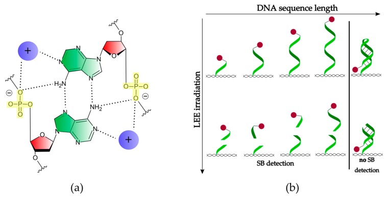
Figure 5.
(a) Scheme of the A-A (green) base pair attached to the deoxyribose sugar unit (red) that builds the poly(A) duplex in the presence of cations (purple). The phosphate groups (yellow) are driven toward the axis of the helix and form two of the total four hydrogen bonds (dotted lines) [25]. (b) Scheme of the conformational change of a single-stranded DNA with increasing length and SB formation. DNA origami is depicted as a pattern in black, the biotin label as a spot in red, and the hydrogen bonding is labeled as a solid line in black.

