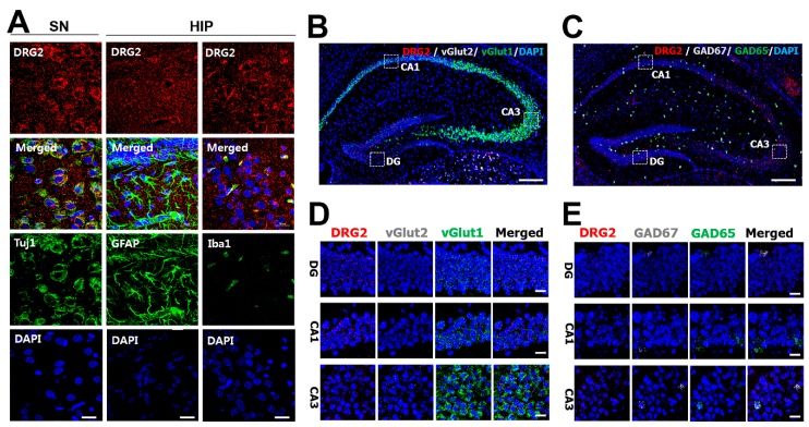
Figure 3.
The expression profiling of DRG2 in the mouse neurons. (A) Immunohistochemical staining of neuronal marker Tuj1, astrocytes marker GFAP, microglia marker Iba1, and DRG2 in neuron cell bodies of substantia nigra (SN) and dentate gyrus (DG) of the hippocampus (HIP). Nuclei were stained with DAPI. Scale bar, 20 μm. (B–E) In situ hybridization analysis of DRG2 expression in mouse hippocampus. (B,C) Hippocampal sections of mouse brain were hybridized with antisense probes against vGlut2, GAD67 (gray dot), vGlut1, GAD65 (green dot), and DRG2 (red dot). Nuclei were stained with DAPI. Scale bar, 200 μm. The boxed regions in images of (B,C) were viewed at a higher magnification in the bottom panels (D,E), respectively. CA1 and CA3, Cornu Ammonis 1 and 3. Scale bar 20 μm.

