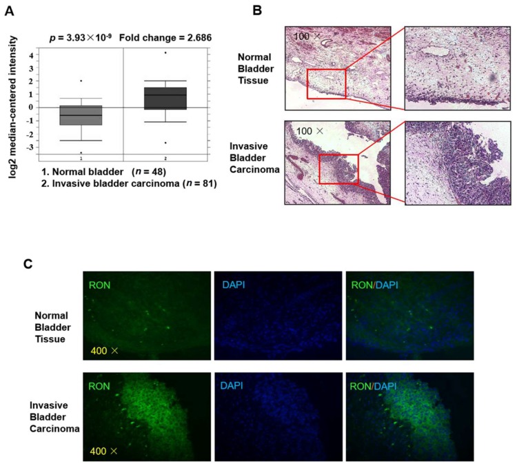
Figure 1.
RON expression is higher in invasive bladder cancer than in normal bladder tissue. (A) The expression level of RON from a clinical sample database (https://www.oncomine.org). (B) H&E staining of normal bladder tissue (upper) and invasive bladder carcinoma (lower) sections. The photographs were taken under a microscope at 100× magnification. (C) Immunofluorescence staining was performed to detect RON protein in normal bladder and invasive bladder carcinoma tissue. RON positive cells were stained green, and the nuclei were stained blue. The photographs were taken with a fluorescence microscope at 400× magnification.

