Figure 4.
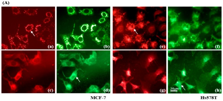
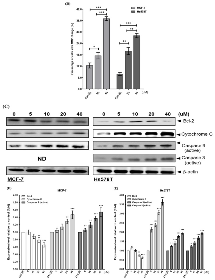
Diosgenin dissipates the mitochondrial membrane potential (∆Ψm) in breast cancer cells. (A) Images of JC-1 fluorescence were taken at 48 h. Red fluorescence indicates aggregates of JC-1 in intact mitochondria, whereas green fluorescence represents the JC-1 monomer in cytosol. MCF-7 and Hs578T cells treated with vehicle are shown in (a,b,e,f), respectively. MCF-7 and Hs578T cells treated with 20 µM diosgenin are shown in (c,d,g,h), respectively. Arrows indicate the translocation of JC-1 from mitochondria (a,e) to cytosol (d,h). (B) Quantitation of the flow cytometric analysis of diosgenin-treated MCF-7 (left panel) and Hs578T (right panel) cells showing loss of the ∆Ψm. Data are shown as means ± SD for three independent experiments. * p < 0.05, ** p < 0.01, and *** p < 0.001 vs. control (Ctrl, 0 µM) cells. (C) Breast cancer cells were treated with various concentrations of diosgenin for 48 h. Western blot analyses with antibodies to apoptosis-relevant proteins. β-actin was used as the loading control. (D,E) Protein levels were determined by Western blot analyses using specific antibodies to the indicated cell cyclins. β-actin was used to normalize the amount of protein in each lane. Data are shown as means ± SD for three independent experiments. * p < 0.05, ** p < 0.01, and *** p < 0.001 vs. control (Ctrl, 0 µM) group.


