Figure 3.
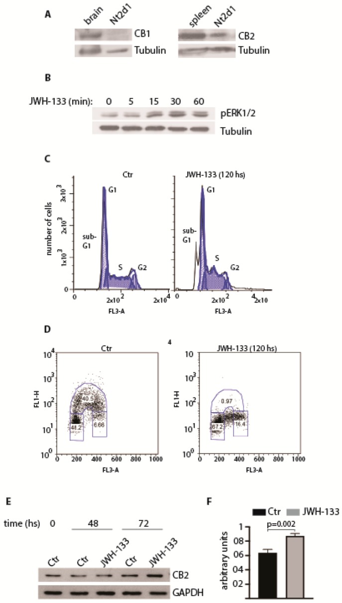
Cannabinoid receptor CB2 in embryonal carcinoma. (A) Expression of CB1 and CB2 receptors in Nt2d1 cell line. Mouse brain and spleen were used as positive controls for the expression of CB1 and CB2, respectively. (B) Analysis of the activation of ERK pathway following the stimulation with 1 µM JWH-133 at the indicated time points. In (A) and (B), tubulin was used as loading control. (C) Cell cycle profile of Nt2d1 cells in untreated and JWH-133 treated cells (1 µM), at the indicated time point. The sub-G1 pick is indicative of nuclear fragmentation. (D) Chronic exposure of Nt2d1 cells at 1 µM JWH-133 causes an arrest at the G1/S phase of the cell cycle. (E) Expression of CB2 receptor following chronic exposure of Nt2d1 cells at 1 µM JWH-133 for the indicated time frames. The expression of glyceraldehyde 3-phosphate dehydrogenase (GAPDH) was used as loading control. (F) Densitometric analysis of CB2 expression treatment of Nt2d1 cells with 1 µM JWH-133 for 72hs. CB2 expression was normalized against the loading control (GAPDH). Data are mean value ± s.d. of three independent experiments. Statistical analysis was performed using a unpaired two-tail Student’s t-test (p < 0.05; Barchi and Grimaldi, unpublished data).

