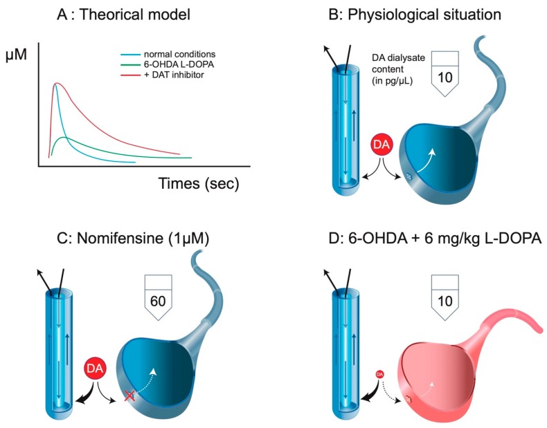
Figure 3.
Clearance and intracerebral microdialysis. (A) Using fast cyclic voltammetric measurements, it is possible to illustrate the clearance of a neurotransmitter like DA. Upon stimulation, the signal (arbitrarily converted to µM) sharply reaches its maximum levels, and the clearance starts. It results in a rapid disappearance of the neurotransmitter from the extracellular space. In the presence of a DA transporter (DAT) blocker, the magnitude of the signal is not necessarily increased, but the rate of disappearance is dramatically reduced. In 6-OHDA conditions, the magnitude of the signal, if any, is lower, but the rate of disappearance is extremely low (no DAT). (B) Striatal DA extracellular levels correspond to a balance between the endogenous clearance and the exogenous clearance due to the microdialysis probe. In classic microdialysis experiments, the basal DA dialysate content in the striatum approximately reaches 10 pg/sample. (C) The local or systemic administration of a DAT blocker results in the increase in DA extracellular levels due to the loss of endogenous clearance and the increase in exogenous clearance (here, by sixfold according to [23]. (D) In 6-OHDA, there is no endogenous clearance of DA. The magnitude of the initial signal explaining the 10 pg obtained in the dialysate after 6–10 mg/kg L-DOPA is necessarily very low. Adapted from [20].

