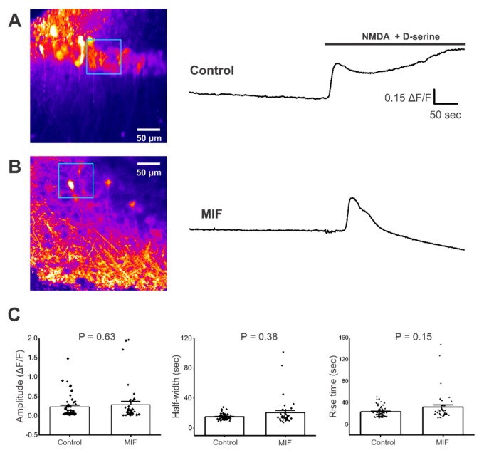
Figure 2.
NMDA + D-serine response in CA1 neuronal somata of hippocampal brain slices. All experiments were conducted in aCSF with 0 Mg2+ and 10 μM Bicuculline. (A) Left, Representative image of GCaMP expression in the CA1 layer of hippocampal brain slices from control mice (blue square represents ROI). Right, representative trace from ROI, with and without NMDA + D-serine application; (B) Left, Representative image of GCaMP expression in the CA1 layer of hippocampal brain slices from MIF mice (blue square represents ROI). Right, representative trace from ROI with and without NMDA + D-serine application; (C) Average amplitude, half-width and rise time data of NMDA + D-serine response from two to four slices each, from eight control mice and four MIF-treated mice. p values based on Mann-Whitney test are shown for graphs in C.

