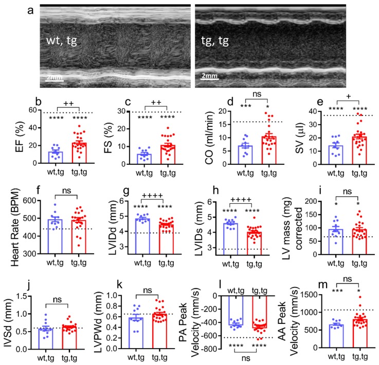
Figure 4.
Cardiac corin-Tg(i) overexpression improves systolic function in female mice with DCM. (a) Representative two-dimensional guided left ventricular short axis M-mode images, (b) ejection fraction (EF), (c) fraction shortening (FS), (d) cardiac output (CO), (e) stroke volume (SV), (f) heart rate (beats per minute), (g) left ventricular internal diameter in diastole (LVIDd), (h) left ventricular internal diameter in systole (LVIDs), (i) LV mass, (j) interventricular septal wall thickness in diastole (IVSd), (k) left ventricular posterior wall thickness in diastole (LVPWd), (l) pulmonary artery (PA) peak velocity, and (m) ascending aorta (AA) peak velocity in all experimental groups. Experimental groups were mice at 90 days of age: Corin-Tg(i)/DCM (●, tg,tg, n = 21) and corin-WT/DCM (●, wt,tg, n = 10), and dotted line represents normal control values in corin-WT/WT (wt,wt, n = 7). Statistical differences in all panels among three groups were analyzed by one-way ANOVA using Newman–Keuls multiple comparisons test except in (d,i,k,m) where Kruskal–Wallis test using Dunn’s multiple comparisons test were used. Data are represented as mean ± SE; **** p < 0.0001, *** p < 0.001, * p < 0.05 (tg,tg or wt,tg vs. wt,wt); ++++ p < 0.0001, ++ p < 0.01, + p < 0.05 (tg,tg vs. wt,tg); ns = not significant.

