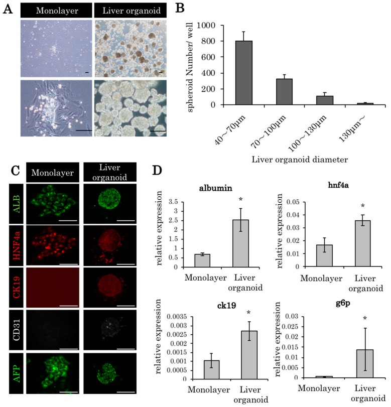
Figure 1.
Liver organoids made from embryonic day 14 (ED14) rat fetal liver cells. (A) Monolayer culture and liver organoids derived from embryonic day (ED) 14 fetal liver cells (culture period, 24 h). Scale bars: 100 μm. (B) The diameters of most of the liver organoids were between 40 and 70 μm. (C) The histopathological findings of the monolayer culture and liver organoids derived from ED14 fetal liver cells (ALB: albumin; HNF4a: hepatocyte nuclear factor 4 alpha; and CK19: cytokeratin 19). Scale bars: 100 μm. (D) Gene expression in monolayer culture and liver organoids 24 h after incubation. In glucose 6 phosphatase (g6p), 96 h culture specimens were used. The relative expression levels of the genes compared to 18s ribosomal RNA are indicated by PCR. * p < 0.05 vs monolayer, Mann–Whitney U test, n = 3.

