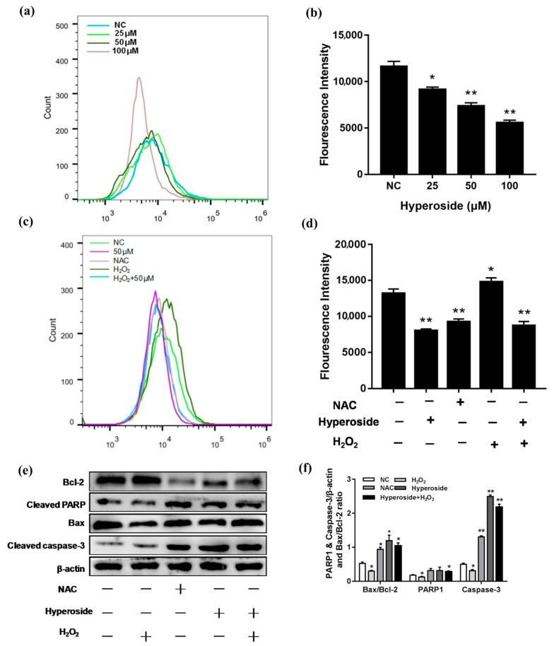
Figure 3.
Hyperoside induces apoptosis by reducing intracellular ROS levels. (a–d) 4T1 cells were stimulated with different concentrations (25, 50 and 100 µM). NAC: Cells were stimulated with 10 mM NAC; H2O2: Cells were stimulated with 25 µM H2O2; hyperoside + H2O2: Cells were co-stimulated with 25 µM H2O2 and 50 µM hyperoside. All these groups were performed for 24 h. The intracellular ROS levels were measured by staining with DCFH-DA (10 mM) for 30 min and then determined by flow cytometry. (e,f) 4T1 cells were stimulated with hyperoside (50 µM), NAC (10 mM), H2O2 (25 µM), and hyperoside (50 µM) + H2O2 (25 µM) for 24 h, respectively. The proteins levels of cleaved caspase-3, Bax, cleaved PARP, and Bcl-2. β-actin were used for normalization. Data are expressed as mean ± SD of three independent experiments. * p < 0.05, ** p < 0.01 (Student’s t-test).

