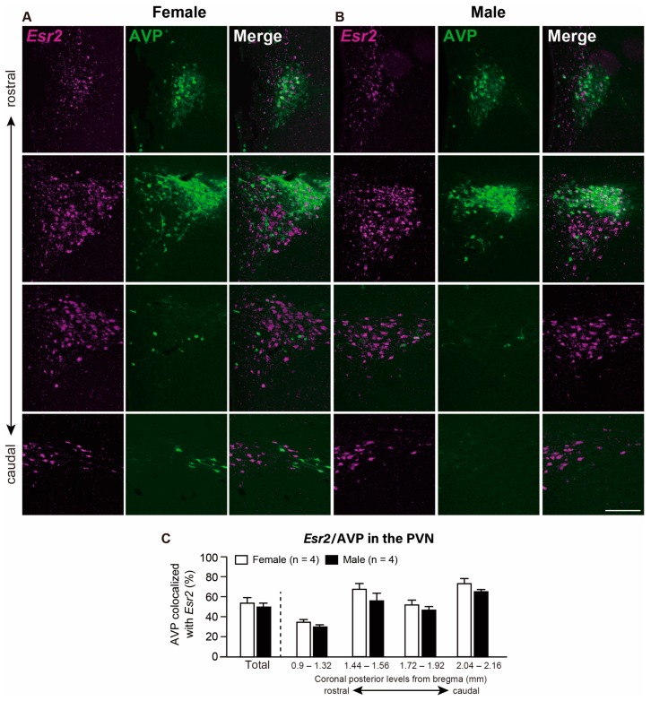
Figure 3.
Co-expression of Esr2 and arginine vasopressin (AVP) in the paraventricular nucleus of the hypothalamus (PVN). Representative photomicrographs show dual labeling of Esr2 mRNA (magenta) and AVP (green) in the PVN of female (A) and male (B) rats. Scale bars indicate 200 µm in A and B. (C) The percentage of AVP-ir cells co-expressed with Esr2 mRNA in the whole PVN and the PVN at different rostro-caudal levels (from bregma −0.9 to −2.16 mm). Values represent the mean ± SEM.

