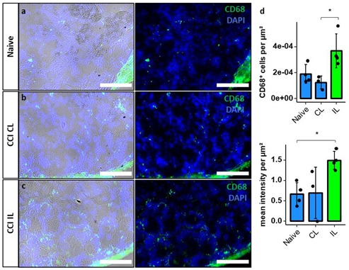
Figure 8.

Increased macrophage invasion in the NRR after CCI. DRGs of naive (a) and contralateral (CL) (b) and ipsilateral (IL) (c) DRGs of Wistar rats 7d after CCI were harvested and stained using DAPI and anti-CD68 antibodies (left: immunofluorescence with brightfield; right: only immunofluorescence). In the NRR, CD68+ cells were counted manually, and signal intensity was measured. Both were quantified per µm2 (d). (n = 3 or 4; CD68+ cells per µm2: CL versus IL, p = 0.024; mean intensity per µm2: Naive versus IL, p = 0.038. Two-way ANOVA, Tukey HSD; scale bars = 100 µm; * p < 0.05).
