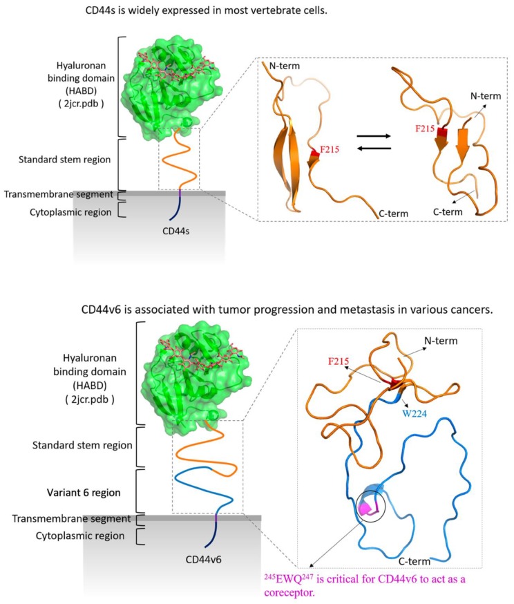
Figure 8.
Schematic for the molecular structures and biological characteristics of CD44s (top) and CD44v6 (bottom). The structure of the HABD (hyaluronan-binding domain) was retrieved from the Protein Data Bank (PDB) (ID: 2JCR), and hyaluronan is shown in sticks. The identified key residue Phe215 is colored red in both CD44s and CD44v6. The standard stem region is colored orange, and the v6 region is colored blue. The amino acids (AAs) 245EWQ247, which are required for CD44v6 to act as the coreceptor for receptor tyrosine kinases (RTKs), including c-Met, Ron, and VEGFR-2, are colored magenta.

