Figure 5.
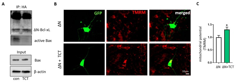
α-TCT protects mitochondrial function in ΔN-Bcl-xL-overexpressing neurons. (A), HEK293T cells were transfected with pME-HA-ΔN-Bcl-xL plasmid. ΔN-Bcl-xL protein was immunoprecipitated with or without α-TCT. ΔN-Bcl-xL protein immunoprecipitated in the presence of α-TCT showed decreased levels of active Bax interaction. Input lanes showed the presence of monomeric Bax in whole cell lysate. Primary hippocampal neurons expressing ΔN-Bcl-xL were treated with or without α-TCT. Neurons treated with α-TCT demonstrated attenuated ΔN-Bcl-xL-mediated loss of mitochondrial potential. TMRM-stained neurons were imaged (B), and TMRM fluorescence intensity was quantified (C) at 6 h after treatment (n = 9–10 cells per group). Scale bar = 10 µm. * p < 0.05, one-way ANOVA.

