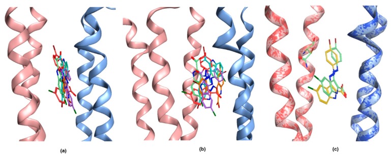
Figure A4.
Different binding poses of MTI163 in the N265S mutant. There is no ligand bound structure with the β1 subunit, thus 6HUP was point mutated for the docking. In this case, five docking poses were found in the top 20 of GoldScore and ChemScore. (a) Front view; (b) Side view; (c) Comparison between the pose displayed in Figure 5 and the most similar pose observed in the N265S mutant. The two bound complexes are superposed (reflected by the two-colored ribbon rendering), the 6HUP (wild type β3) pose and N265 are rendered as light green sticks, the S265 and the corresponding MTI163 pose are rendered as yellow sticks.

