Figure 2.
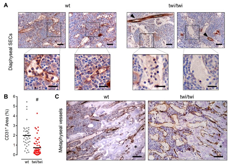
BM diaphyseal twitcher SECs do not express CD31. CD31 immunohistochemical staining of femurs from wt and twitcher mice. (A) CD31 expression in diaphyseal SECs of two wt and two twitcher mice. Arrowheads indicate CD31+ arterioles. Scale bar: 50 μm. Scale bar in magnified fields: 30 μm. (B) Quantification of CD31+ area in femoral histological sections. Dots represent the percentage area occupied by CD31+ vessels in different microscopic fields. Horizontal black bars show the mean value. #, p < 0.001. (C) CD31 expression in metaphyseal vessels. Asterisks indicate the cartilaginous tissue. Scale bar: 100 μm.

