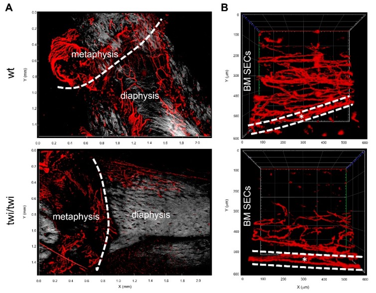
Figure 3.
Two-photon microscopy analysis of CD31 expression in diaphyseal, metaphyseal and cortical bone vessels of twitcher mice. (A) 3D reconstruction of CD31+ vessels (in red) and stromal collagen expression revealed by second harmonic generation (in grey) in diaphysis and metaphysis of wt and twitcher mice (Z = 422 μm and 588 μm for wt and twi/twi specimens, respectively). (B) Cross-sectional view of CD31+ vessels in diaphysis and cortical bone (dashed white lines) of wt and twitcher mice (Z = 294 μm and 352 μm, respectively).

