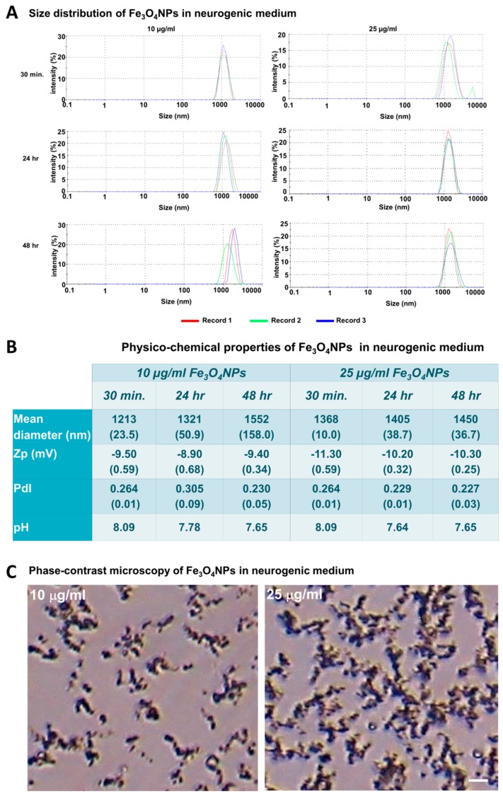
Figure 6.
Physico-chemical characteristics of the Fe3O4NPs in mesenchymal stem cell neurogenic differentiation medium. (A) Size distribution obtained from dynamic light scattering measurements of Fe3O4NPs at concentrations of 10 and 25 μg/mL in mesenchymal stem cell neurogenic differentiation medium after 30 min, 24 and 48 h. (B) Physico-chemical properties of the Fe3O4NPs in mesenchymal stem cell neurogenic differentiation medium. (C) Phase-contrast micrographs of Fe3O4NPs in neurogenic medium at 10 and 25 μg/mL after 48 h: aggregations/agglomerations of the Fe3O4NPs were observed as brownish sediments, which increased as function of the concentration. Scale bar: 20 μm.

