Figure 1.

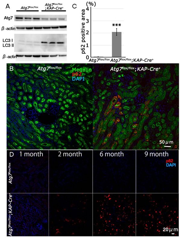
Impairment of autophagy in Atg7-deficient renal proximal tubular cells. (A) Decrease of Atg7 and increase of LC3-I in 2-month-old Atg7flox/flox;KAP-Cre+ mouse kidney. Atg7 and LC3 Western blot (LC3-I/unconjugated LC3 and LC3-II/lipidated LC3) using whole kidney lysate of Atg7flox/flox;KAP-Cre+ mice. As a control, Atg7flox/flox mouse kidney was employed. The intensity of each band of Atg7 and β-actin was estimated by densitometry. The ratio of Atg7 to β-actin was calculated. The amount of Atg7-positive signals in the Atg7flox/flox;KAP-Cre+ mice kidney was about 63% lower than that in the Atg7flox/flox mice kidney (n = 3). Note that LC3-I was increased in the Atg7flox/flox;KAP-Cre+ mouse kidney. (B) The massive accumulation of p62 in kidneys of 2-month old Atg7flox/flox;KAP-Cre+ mouse. The cortico-medullary region of each kidney in 2 month-old Atg7flox/flox;KAP-Cre+ mouse and Atg7flox/flox mouse was recognized with anti-p62 antibody (red). As a marker of renal proximal tubular cells, megalin (green) was employed. Nuclei were stained with 4′,6-diamidino-2-phenylindole (DAPI; blue). (C) Quantification of p62 positive area of 2-month-old Atg7flox/flox;KAP-Cre+ (n = 5) and Atg7flox/flox kidney (n = 4, *** p < 0.01). Data in graphs are expressed as the mean ± SEM. Statistical analyses were performed using a student’s t-test. (D) Age-dependent accumulation of p62 (red) in the Atg7flox/flox;KAP-Cre+ mouse kidney in 1-, 2-, 6-, and 9-month-old Atg7flox/flox;KAP-Cre+ (lower) and Atg7flox/flox (upper) mouse kidney.
