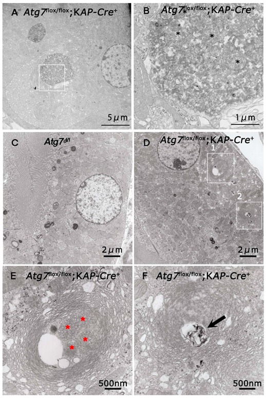
Figure 5.

Electron microscopic analyses of Atg7 deficient renal proximal tubular cells. The renal proximal tubular cells of 2-month-old Atg7flox/flox;KAP-Cre+ (A,B,D–F) and Atg7flox/flox (C) mice were investigated with a transmission electron microscopy. (B) is higher magnification of area. Black asterisks indicated degenerated mitochondria. (A) (white box). (E,F) are higher magnification of areas (white boxes 1 and 2, respectively) in (D). Asterisks in (E) indicated peroxisome-like structures in the multi-lamellar bodies. Arrows indicate lysosome like structures in the multi-lamellar bodies.
