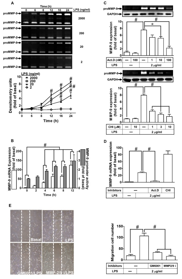
Figure 1.
Lipopolysaccharide (LPS) induced metalloproteinase-9 (MMP-9) expression and cell migration in rat brain astrocytes (RBA-1) cells. (A) Cells were incubated with various concentrations of LPS (2, 0.2, 0.02, and 0.002 μg/mL) for the indicated time intervals (0, 6, 8, 12, 16, and 24 h). The levels of MMP-9 were determined by gelatin zymography. (B) Cells were incubated with LPS (2 μg/mL) for the indicated time intervals (0, 2, 4, 6, 8, and 12 h). The levels of MMP-9 mRNA and promoter activity were analyzed by real-time PCR and promoter activity assay, respectively. (C) Cells were pretreated with actinomycin D (Act. D; 0.001, 0.01, and 0.1 μM) or cycloheximide (CHI; 1, 3, and 10 μM) for 1 h, and then incubated with LPS (2 μg/mL) for 24 h. The levels of MMP-9 were determined by gelatin zymography. The glyceraldehyde-3-phosphate dehydrogenase (GAPDH) level of cell lysates was assayed by western blot. (D) Cells were pretreated with Act. D (0.1 μM) or CHI (10 μM) for 1 h, and then incubated with LPS (2 μg/mL) for 4 h. The level of MMP-9 mRNA expression was determined by real-time PCR. (E) Cells were pretreated with GM6001 (1 μM) or MMP2/9 inhibitor (MMP2/9i, 1 μM) for 1 h and then incubated with LPS (2 μg/mL) for 48 h, and cell migration was assayed (magnification = 40×). Data are expressed as mean ± SEM of three independent experiments. * p < 0.05; # p < 0.01, as compared with the cells exposed to vehicle or LPS as indicated.

