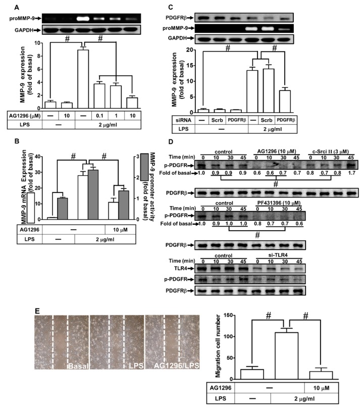
Figure 5.
Platelet-derived growth factor receptor (PDGFR)β was involved in LPS-induced MMP-9 expression and cell migration. (A) RBA-1 cells were pretreated with AG1296 (0.1, 1, and 10 μM) for 1 h, and then incubated with LPS (2 μg/mL) for 24 h. The levels of MMP-9 were determined by gelatin zymography. The GAPDH level of cell lysates was assayed by western blot. (B) Cells were pretreated with AG1296 (10 μM) for 1 h, and then incubated with LPS (2 μg/mL) for 4 h for mRNA expression or 6 h for promoter activity. The mRNA expression and promoter activity of MMP-9 were determined by real-time PCR and promoter assay, respectively. (C) Cells were transfected with scrambled (Scrb) or PDGFRβ siRNA, and then incubated with LPS (2 μg/ml) for 24 h. The medium and cell lysates were collected to respectively determine the levels of MMP-9 by gelatin zymography, and the levels of GAPDH and PDGFRβ by western blotting. (D) Cells were pretreated with or without AG1296 (10 μM), PF431396 (10 μM), or c-Src inhibitor II (3 μM) for 1 h, or separately transfected with TLR4 siRNA, and then challenged with LPS (2 μg/mL) for the indicated time intervals (0, 10, 30, and 45 min). The phosphorylation of PDGFRβ was determined by western blotting. (E) Cells were pretreated with AG1296 (10 μM) for 1 h, and then incubated with LPS (2 μg/mL) for 48 h. The number of cell migrations was determined (magnification = 40×). Data are expressed as mean ± SEM of three independent experiments. # p < 0.01 as compared with the cells exposed to vehicle or LPS, as indicated.

