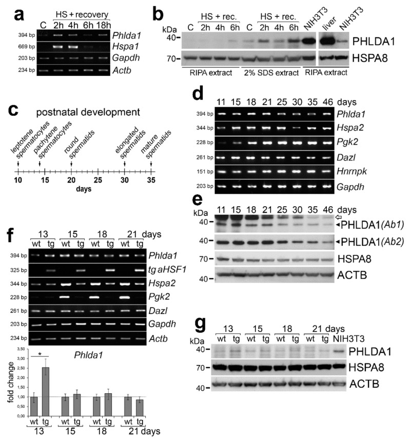
Figure 1.
Expression of pleckstrin-homology-like domain family A, member 1 (PHLDA1) in mouse testes. (a) Transcripts of Phlda1 and reference genes analyzed by RT-PCR in adult testes after heat shock performed in vivo and indicated recovery time. C—control, physiological temperature; HS—heat shock. (b) PHLDA1 protein level analyzed by western blot in testes of mice subjected to heat shock and indicated recovery time. HSPA8 was used as loading control; proteins were extracted with either RIPA or 2% SDS buffer. (c) Time-line of the appearance of different spermatogenic cells during the mouse postnatal development. (d) Transcripts of Phlda1 and reference genes analyzed by RT-PCR in testes of 11–46-day-old animals. (e) PHLDA1 protein level analyzed by western blot in testes of 11–46-day-old animals. ACTB was used as loading control; two anti-PHLDA1 antibodies (Ab1 and Ab2) were used; heavy chain IgG detected by the secondary anti-mouse antibody is marked with an arrow. (f) Transcripts of Phlda1 and reference genes analyzed by RT-PCR in testes of wild-type (wt) and aHSF1 transgenic (tg) mice at 13th, 15th, 18th, and 21st day of postnatal development (upper panel); fold change in Phlda1 expression quantified by RT-qPCR in testes of tg mice compared to wt mice of the same age (bottom panel; marked are minimum and maximum values). Asterisk indicates the statistical significance of differences (* p < 0.05). (g) PHLDA1 protein level analyzed by western blot in testes of wild-type (wt) and aHSF1 transgenic (tg) mice. ACTB and HSPA8 were used as loading controls.

