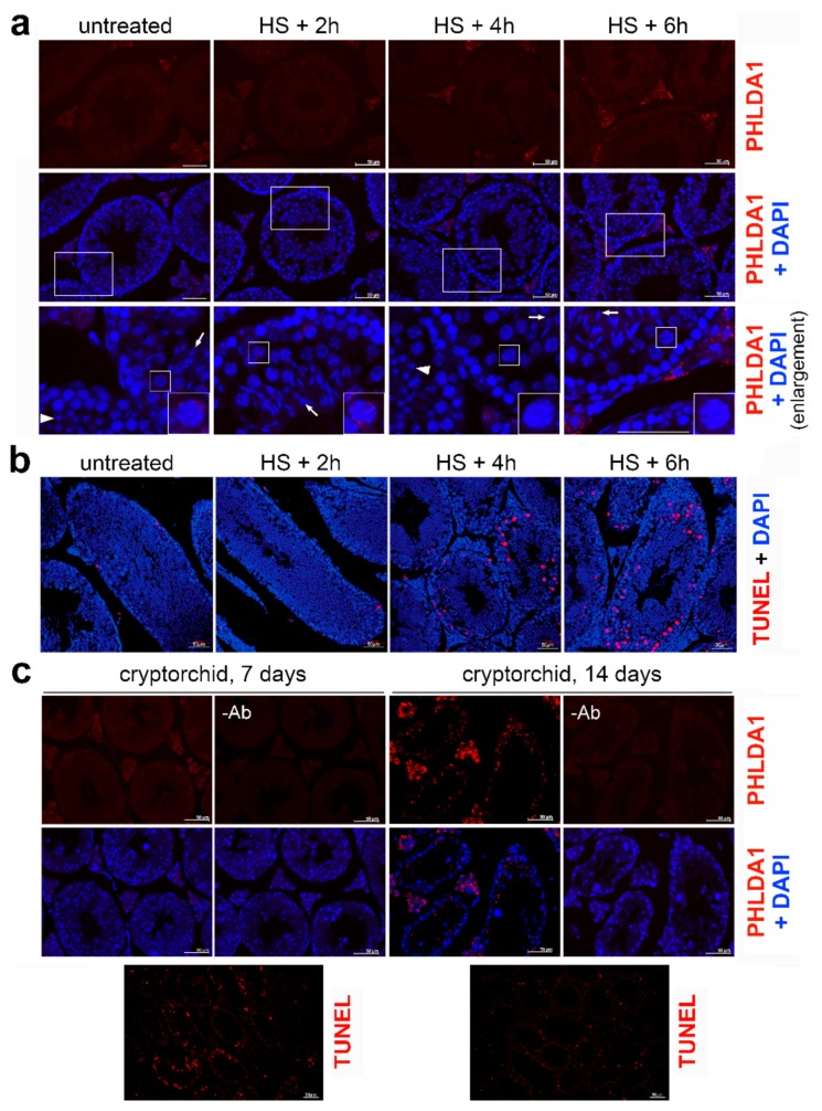
Figure 2.
Localization of PHLDA1 in mouse testes. (a) Detection of PHLDA1 by immunofluorescence (using Ab2, red; DNA stained with DAPI, blue) in testes of a control mouse and subjected to heat shock after 2, 4, and 6 h of recovery. Enlargement of the marked areas is shown in the bottom panel; round spermatids and condensing spermatids are marked with arrowhead and arrows, respectively. Representative pachytene spermatocytes (in squares) are further enlarged in the lower corners. (b) Detection of apoptotic DNA breaks (by TUNEL assay, red; DNA stained with DAPI, blue) in seminiferous tubules of untreated mice and after heat shock in vivo and indicated recovery time (2–6 h). (c) Detection of PHLDA1 by immunofluorescence (red; DNA stained with DAPI, blue) in cryptorchid testes 7 and 14 days after surgery (upper panel) and detection of apoptotic DNA breaks (by TUNEL assay, red) in corresponding tissues (bottom panel). Scale bar—50 µm.

