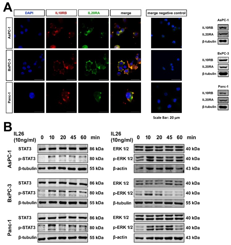
Figure 4.
PDAC cell lines. (A) Laser scan microscopy and Western blotting of the PDAC cell lines AsPC-1, BxPC-3, and Panc-1 incubated with anti-IL10RB and anti-IL20RA antibodies shows expression and colocalization of both receptors in all three cell lines. (B) Western blotting of PDAC cells shows IL26 induced phosphorylation of STAT3 (left panel) and ERK1/2 (right panel) in a time-dependent manner.

