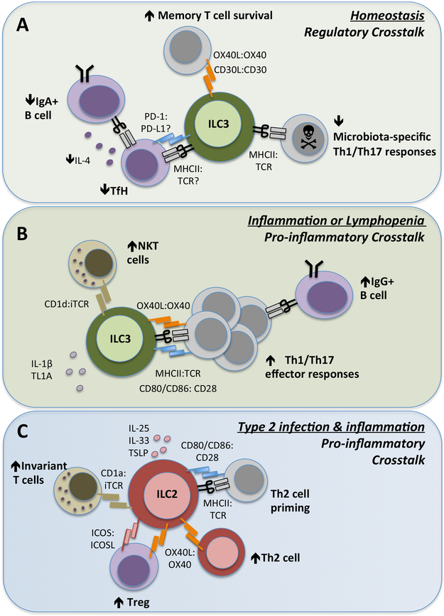
Figure 4 ∣. ILCs control adaptive immunity through direct cellular interactions.
ILC subsets, in particular ILC2s and ILC3s, express a range of surface molecules that facilitate direct cell–cell interactions and modulation of T cell responses in health and disease. a) ILC3s constitutively express MHC class II molecules in intestinal-draining lymphoid tissues and the colon at homeostasis. In this context, ILC3s lack surface co-stimulatory molecules and present antigens to microbiota-specific CD4+ T cells, resulting in the induction of cell death. Similarly, interactions with TFH cells within the mesenteric lymph node limits TFH cell-derived IL-4 and subsequent class-switching of mucosal B cells to produce IgA. ILC3 also express PD-L1, which may interact with PD-1 on TFH cells, but this has yet to be extensively explored. ILC3 expression of non-canonical co-stimulatory molecules such as OX40L and CD30L can support memory T cell responses, although it is unknown whether this also requires MHC class II-dependent antigen presentation. b) By contrast, in the context of a maladapted immune system, such as in lymphopenia or chronic inflammation, the antigen-presenting function of ILC3s can change markedly. In response to pro-inflammatory cytokines such as IL-1β and TL1A, ILC3s upregulate expression of co-stimulatory molecules including CD80, CD86 and OX40L and promote inflammatory T cell effector responses. c) Upon activation by IL-25 and/or IL-33 (derived from epithelial cells, stromal cells and myeloid cells), ILC2s upregulate expression of MHC class II and co-stimulatory molecules such as CD80 and CD86, which facilitate direct interactions with naïve T cells and promote priming of TH2 cells in the context of allergic airway inflammation or infection. In addition, ILC2 expression of OX40L promotes the proliferation and effector responses of TH2 cells and further supports expansion of OX40+ Treg cells. ILC2s further enhance Treg cell responses through provision of surface ICOS, which binds ICOSL expressed on Treg cells.

