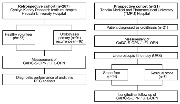
Figure 1.
Flow diagram of the retrospective and prospective observational studies. The retrospective study enrolled 110 patients diagnosed with urinary calculi and 157 healthy volunteers who received health checks at Oyokyo Kidney Research Institute Hospital between June 2015 and August 2018. All urine samples were collected before stone treatment, and then the urine protein concentration was adjusted to 2 mg/mL followed by storage at −80 °C until use. A prospective cohort enrolled 21 patients with patients who were diagnosed with urinary calculi at Tohoku Medical and Pharmaceutical University Hospital in Sendai Japan between April 2018 and May 2019. Urine was collected prospectively in a patient diagnosed with urinary calculi during stone treatment. We divided the patients into two groups during stone treatment: the group without presence of stones after URS (stone-free group, n = 14) and the group with presence of stone after URS (residual-stone group, n = 7). As a diagnosis of urolithiasis, computed tomography was used for detecting the presence or absence of calculus. The definition of the presence of urolithiasis was based on CT imaging of a patient with a urinary calculus > 4 mm. The exclusion criteria were patients with renal atrophy, urinary catheter, and renal failure. Gal3C-S-OPN and uFL-OPN concentration were measured.

