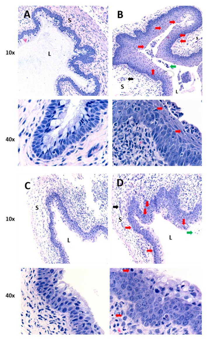
Figure 2.
Histology of vaginal tissues from mice treated with curcumin nanoparticles by intravaginal delivery and challenged with CpG-ODN. All mice were depo-treated (n = 3–4 per group). (A) Mice received water with no CpG-ODN (negative control). All other groups (B–D) received IVAG inoculation with 30 µg CpG-ODN 2 h after primary treatment with: (B) water (positive control), (C) 0.5 mg of curcumin in nanoparticles IVAG, or (D) vehicle-only nanoparticles IVAG. Images are representative from vaginal tissue of 1 animal per treatment group, with magnifications of 10× and 40×. L denotes vaginal lumen and S denotes the vagina submucosa. Red arrows indicate thickened vaginal epithelium and/or inflammatory cell infiltration in the epithelium and submucosal tissue. Black arrows indicate inflammatory cell infiltrates in the blood vessels. Green arrows indicate inflammatory cells in the luminal space.

