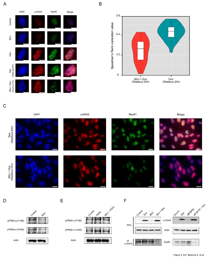
Figure 4.
Inhibition of FGF-signaling impairs RAD51 loading to DNA DSBs: (A) GIST T-1R cells were pretreated with Dimethyl Sulfoxide (DMSO) (control) or BGJ398 (1 µM) for 48 h prior doxorubicin treatment (0.25 µg/mL) and 4 h later processed for immunofluorescence using Rad51 and γ-H2AX. DAPI staining was used to outline the nucleus (Scale bars = 10 μm). (B) Graph illustrating the distributions of Spearman’s rank correlation values between GIST T1-R cells treated with Dox alone (mean = 0.429) or in presence of BGJ398 (mean = 0.245). p value = 9.816e-10 (t-test). (C) Immunofluorescence staining for H2AX and Rad51 was performed on Dox-treated GIST cells after DMSO (control) or BGJ398 treatment. A wider field of cells is shown (scale bars = 20 μm). BGJ398 inhibits FGFR-signaling in IM-resistant and naïve GIST cells: (D) BGJ398 abolishes the FGFR-signaling in IM-resistant GIST T-1R cells. The cells were treated with DMSO (control) and BGJ398 (1 μM) for 72 h and were further subjected to immunoblotting with phosphorylated forms of FRS-2 antibodies. (E) BGJ398 abrogates FGF-2-induced activation of FGFR-signaling in IM-resistant GIST T-1 cells. The cells were treated with DMSO (control), FGF-2 (100 ng/mL) alone or in presence of BGJ398 (1 μM) for 72 h and were further subjected to immunoblotting with phosphorylated forms of FRS-2 antibodies. (F) GIST T-1R cell lysates were immunoprecipitated with γ-H2AX Abs and immunoblotted with Rad51 Abs to demonstrate endogenous complex formation. A whole cell lysate (WCL) was included. Actin was used as a loading control. Inhibition of FGF-signaling was achieved by using BGJ398, a selective FGFR 1-4 inhibitor (left), or siRNA against FGFR2 (right). GIST T-1R cells were pre-treated with BGJ398 1 µM (left) or transfected with siFGFR2 (right) for 48 h before Dox treatment (1 µg/mL for 24 h).

