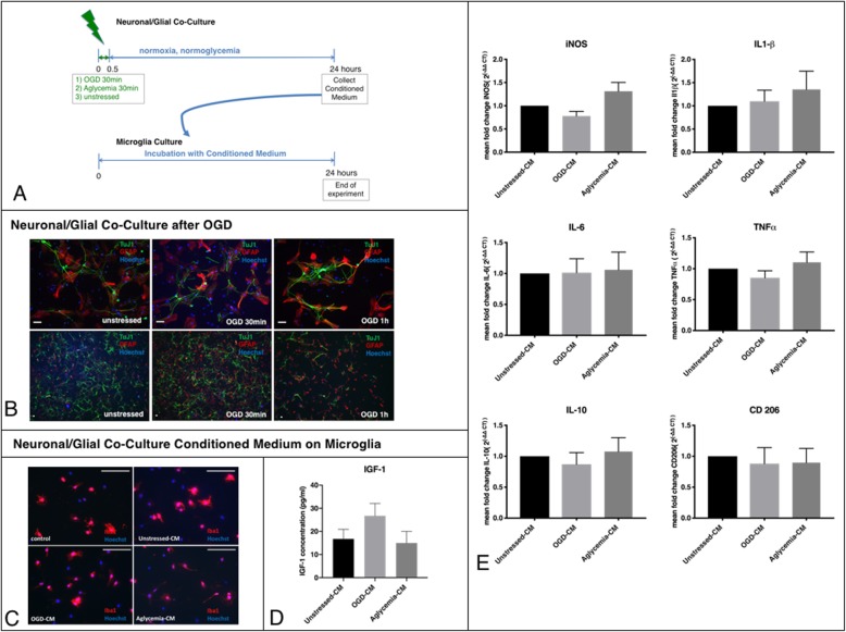
Fig. 3.
Effects of neuronal/glial co-cultures exposed to metabolic stress on microglia. a Experimental timeline. Neuronal/glial co-cultures differentiated from primary neural stem cells were exposed to metabolic stress, either as OGD or as aglycemia, for 30 min, before they were allowed to recover in regular culture medium for 24 h. The supernatant of preconditioned neuronal/glial co-cultures (conditioned medium, CM) was used for further experiments. Unstressed neuronal/glial co-cultures served as control. The CM of all conditions was incubated with microglia for 24 h. Then the microglia were used for further experiments. b Representative immunocytochemical images of neuronal/glial co-cultures after OGD. Tuj1+ neurons (green) and GFAP+ astrocytes (red) were co-stained with a nuclear marker (Hoechst; blue) in either unstressed microglia (left panel), microglia after 30 min of OGD (middle panel) or microglia after 1 h of OGD (right panel; scale bars = 50 μm), showing less viable cells after 1 h of OGD. c Representative immunocytochemical images of microglia incubated with the CM of pre-conditioned neuronal/glial co-cultures. Iba1+ microglia (red) were co-stained with a nuclear marker (Hoechst; blue) in either untreated microglia (upper left panel), microglia with unstressed-CM (upper right panel), or microglia with OGD-CM (lower left panel), or aglycemia-CM, respectively (lower right panel; scale bars = 50 μm), showing no changes in morphology under these different conditions. d An IGF1-ELISA was used to quantify IGF1 release in the microglia supernatant photometrically. There were no significant changes in IGF1 release between untreated control microglia and microglia incubated with either unstressed-CM and OGD-CM, or aglycemia-CM (values displayed as means ± SEM of three independent experiments with n = 12/each condition; one-way ANOVA). e Q-PCR revealed that neuronal/glial co-cultures exposed to metabolic stress did not affect the expression of either M1 or M2 markers on microglia: iNOS, TNFα, IL-6, IL1-β, IL-10, and CD 206 were unchanged compared to microglia incubated with non-preconditioned medium (values displayed as means ± SEM of three independent experiments with n = 12/ each condition, one-way ANOVA). Each Q-PCR sample was normalized to RPL13a as the reference gene, and mRNA levels were normalized to the endogenous RPL13a expression

