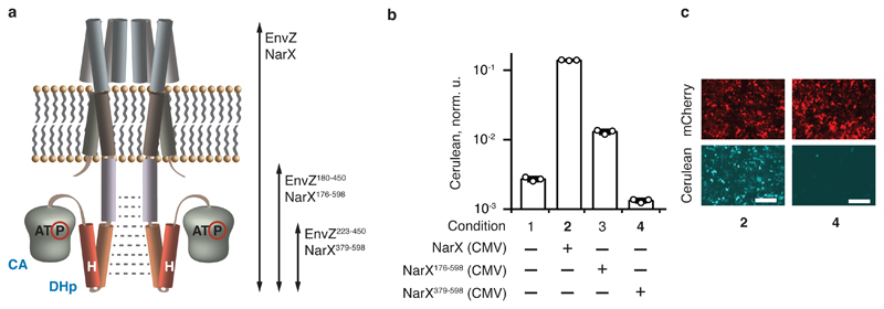
Fig. 1. Identification of HK domains with reduced intrinsic signaling.
a, A representation of an HK receptor in a cell membrane. CA domain with bound ATP and DHp domain containing the phosphorylatable histidine (H) are shown. Dimerization is indicated with dotted gray lines. The arrows span the various tested domains (see also Supplementary Fig. 2). b, Signaling capacity of truncated NarX. The bars show Cerulean levels in cells coexpressing NarL and an indicated NarX variant in the presence of NarL-inducible Cerulean reporter. Cerulean expression is normalized to the transfection control and shown as mean ± SD of independent biological triplicates. The circles indicate individual measurements. c, Microscopy images of HEK293 cells for conditions shown in bold in panel b. The top and the bottom rows show, respectively, the expression of constitutive mCherry transfection control (red), and pathway-induced Cerulean reporter (cyan) in the same transfection. The numbers correspond to the conditions in b. The white scale bar is 200 μm. The DNA constructs are described in Supplementary Fig. 1. The results were reproduced at least once in an independent experiment.

