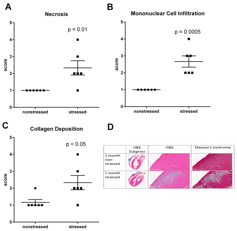
Fig. 3. Stress induces myocardial lesions characterized by fibrosis, necrosis, and mononuclear cell infiltration.
Hearts from stressed and nonstressed rats were evaluated by H&E and Masson’s trichrome staining. Hearts from stressed rats developed multifocal lesions characterized by necrosis (A,D), infiltration by mononuclear immune cells (B,D), and collagen deposition (C,D). Data represent the mean ± S.E.M. of 6 animals. Representative photographs of heart sections are shown in panel D. Blue circles in photographs of subgross heart images indicate heart regions that were photographed at higher (15.8 X) magnification. Scale bars in panel D represent 200 µm.

