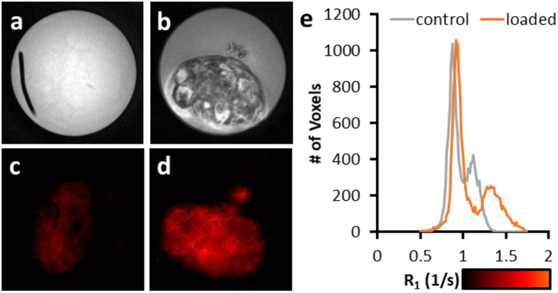
Figure 4:
A comparison of T1-weighted images from porcine ovaries perfused with VS55 (a) and IONPs (PMG-300; 429 mMFe) (b). The IONP perfused ovary allows for the visualization of the follicles of the ovary. Changes in R1 can be observed from the porcine ovaries perfused with VS55 (c) and IONPs (d). The R1 increases by 0.21 s−1 between the VS55 and IONP perfused ovary (e).

