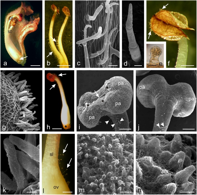
Fig. 2.
Micromorphology of O. picridis stamens and pistils. a Longitudinal section of the flower with visible stamens (st), pistil (pi), and nectary (arrow). b Stamens with visible non-glandular trichomes (arrows) on their filaments. c Non-glandular trichomes on the stamen filaments. d Three-cellular non-glandular trichome. e Glandular trichome on the stamen filament near the anther. f Stamen filament with visible non-glandular trichomes (arrows) on anthers. g, k Non-glandular trichomes located along anther sutures. k Note the massive cuticular striae on the trichome surface. h Pistil with visible nectary (asterisk) and glandular trichomes (arrows) near stigma (st). i, j Stigma covered with papillae (pa) with a visible slit (arrows) and viscous substance (asterisks) on the surface. Note glandular trichomes (arrowheads) on the style surface. i Front view. j Back view. l Fragment of the pistil style (sl) and ovary (ov) with non-glandular trichomes (arrows). m, n Papillae on the stigma surface covered with numerous threads of viscous substance. Scale bars = 2 mm (a, b, h), 500 μm (f, l), 250 μm (i, j), 100 μm (c, g, m), 30 μm (d), 20 μm (e, k), 10 μm (n)

