Fig. 3.
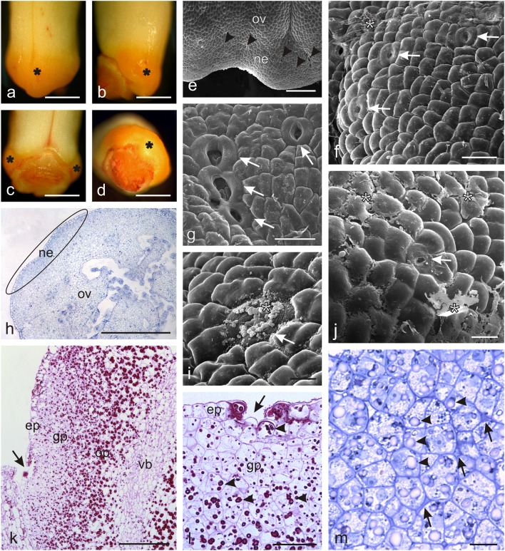
Microstructure of O. picridis nectaries. a–d Gynoecial nectary (asterisks) in O. picridis flowers. a View of the side of the lower lip. b Lateral view. c View of the side of the upper lip. d Top view. e Fragment of the ovary and nectary surface with nectarostomata (arrowheads). f, g, i, j Fragments of the nectary surface with nectarostomata (arrows). Note the dried secretion (asterisks) on the nectary surface. h Cross-section of an ovary with a nectary (elipsa). k Fragment of the longitudinal section of an ovary with a nectary (PAS). Note numerous starch grains in the ovary and glandular parenchyma cells. Vascular bundle visible in the ovary wall (vb). Nectarostoma visible in the nectary epidermis (arrow). l, m Fragments of longitudinal sections of nectariferous tissue. l Visible epidermis with nectarostoma (arrow) and glandular parenchyma with starch grains (arrowheads) (PAS). m Different-shaped glandular parenchyma cells with visible starch grains (arrowheads) and intracuticular spaces with dark content (arrows); ov ovary, ne nectary, ep epidermis, gp glandular parenchyma, op ovary parenchyma. Scale bars = 1 mm (a–d, h), 200 μm (e, k), 50 μm (f, g, j, l), 20 μm (i, m)

