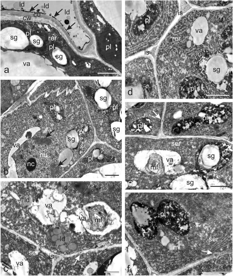
Fig. 4.
Ultrastructural traits of the O. picridis nectary. a Fragment of secretory epidermis cells. Visible dense cytoplasm with plastids filled with starch grains and dense osmiophilic material content, rer, lipid droplets, and large vacuoles. Note the thin osmiophilic film (arrows) on the cuticle surface (cu). b–f Cells of glandular parenchyma with dense cytoplasm, polymorphic plastids, small vacuoles, and thin cell walls. b Note the large nucleus with a conspicuous nucleolus and dark areas with heterochromatin (arrows) and plastids with starch grains and areas with transparent and osmophilic content. c Visible numerous lipid droplets, well-developed ser, vacuoles with myelin-like multilamellar figures, plasmodesmata (arrow), and intercellular space with dense content. d Note numerous rough endoplasmic reticulum profiles forming closely packed strands, plastids with starch grains, and intercellular space with dense content. e Visible plastids with large starch grains, smooth endoplasmic reticulum profiles, plasmodesmata (arrow) in the cell wall, and myelin-like multilamellar figures in the vacuole. f Note the dividing plastid, ser profiles, mitochondria, and plasmodesmata (arrow); pl plastids, sg starch grains, va vacuoles, ld lipid droplets, ser smooth endoplasmic reticulum, rer rough endoplasmic reticulum, mi mitochondria, mf myelin-like multilamellar figures, is intercellular spaces. Scale bars = 2 μm (a, b, d–f), 1 μm (c)

