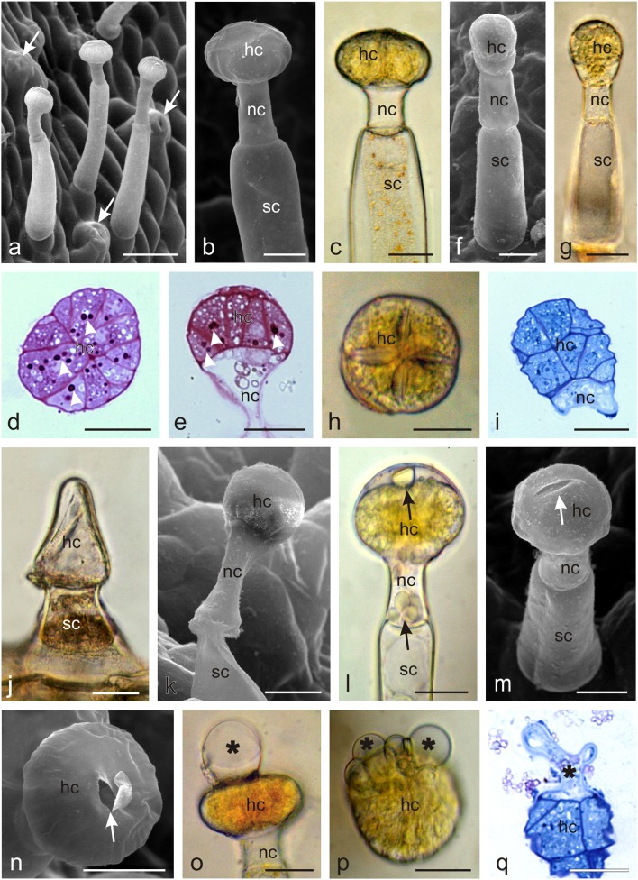
Fig. 5.
Microstructure of glandular trichomes from the abaxial surface of an O. picridis sepal and petal. a Visible capitate glandular trichomes and stomata (arrows). b–e Glandular trichomes type I with a multicellular secretory head formed by cells arranged in a circle. d, e Cross (d) and longitudinal (e) sections of glandular trichomes type I. Note the dense cytoplasm with stained starch grains (arrowheads) (PAS reaction) and fine vacuoles. f–i Glandular trichomes type II with a two-layered head. h Trichome type II head on the upper side. i Longitudinal section of trichome head type II. Visible dense cytoplasm with a large nucleus and small vacuoles. j Conoidal trichomes with the characteristic long conical glandular cell and a short bicellular stalk. k Ageing trichome type I with a narrowed neck and stalk cells. l Droplets of secretion (arrows) visible in the subcuticular space and in the neck cell. m, n Note the ruptures (arrows) in the cuticle on the trichome apex. o–q Visible trichome heads with secretion (asterisks) exuded from ruptures in the cuticle. o, q lateral view. p Top view.; hc head cells, nc neck cells, sc stalk cells. Scale bars = 100 μm (a), 50 μm (g, j), 30 μm (b–f, h, i, k–q)

