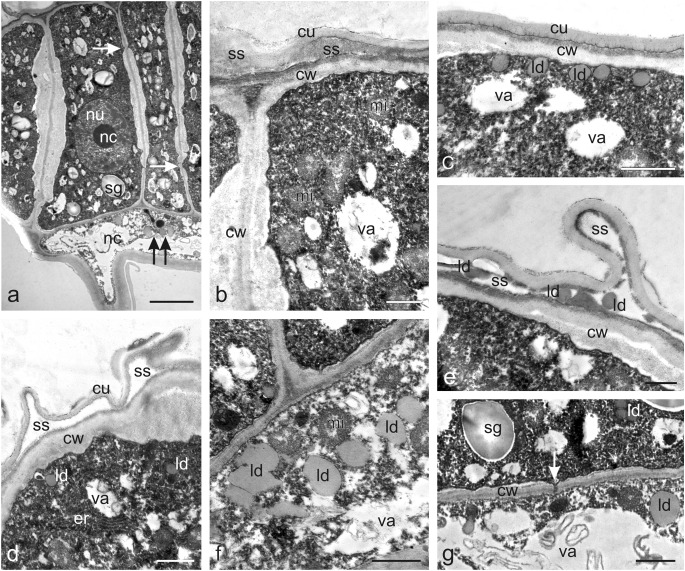
Fig. 6.
Ultrastructural traits of capitate glandular trichomes (type I) on the O. picridis sepal. a Fragment of longitudinal section of trichome head and neck cells. Note the dense cytoplasm with starch grains in the plastids, the large nucleus with a prominent nucleolus, small vacuoles, and plasmodesmata (arrows) in secretory cell walls. Lipid droplets visible in the neck cell (two arrows). b Apical part of the secretory cells of the trichome head with numerous mitochondria and protruding cuticle forming subcuticular space. c Apical fragment of a glandular head cell with numerous lipid droplets visible under the outer cell wall. d Apical fragment of a glandular head cell with visible endoplasmic reticulum profiles and protruding cuticle forming subcuticular space. e Lipid droplets visible in the subcuticular space. f Fragment of a neck cell with visible mitochondria and lipid droplets. g Visible plasmodesmata (arrow) connecting the head cell and the neck cell. b, d, f, g Some membranous remnants visible in vacuoles. hc head cells, nc neck cells, sg starch grains, nu nucleus, nc nucleolus, va vacuoles, cw cell walls, mi mitochondria, cu cuticle, ss subcuticular space, ld lipid droplets, er endoplasmic reticulum. Scale bars = 5 μm (a), 2 μm (g), 1 μm (b–f)

