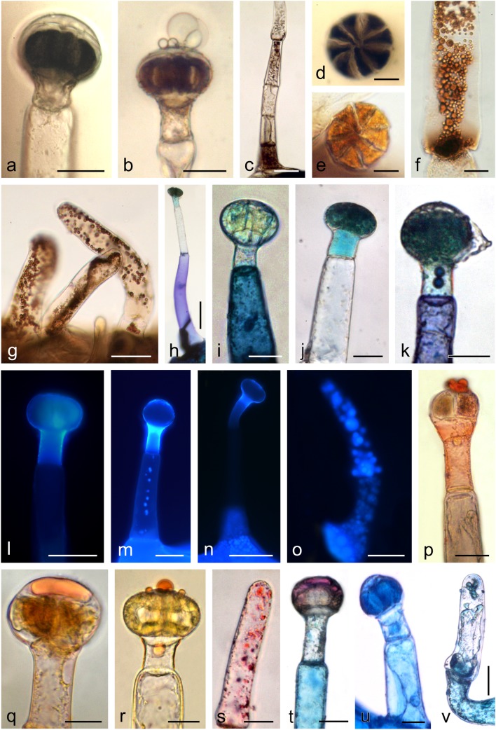
Fig. 7.
Histochemical and fluorescence tests of glandular (a, b, d–f, h–n, p–r, t, u) and non-glandular trichomes (c, g, o, s, v) in O. picridis. a–d Staining of polyphenols with FeCl3. e–g Staining of tannins with potassium dichromate. h–k Staining of polyphenols with Toluidine Blue O. l–o Fluorescence of flavonoids with aluminium chloride (l, m) and magnesium acetate (n, o) fluochromes in the Cy5 filter set. p–s Staining of lipids with Sudan Red B (p, s) and Sudan IV (q, r). t–v Staining of neutral (t) and acidic (u, v) lipids with Nile Blue. Scale bars = 100 μm (n), 50 μm (c, g, h, l, m, o), 30 μm (a, b, i–k, p, s–u), 20 μm (d–f, q, r, v)

