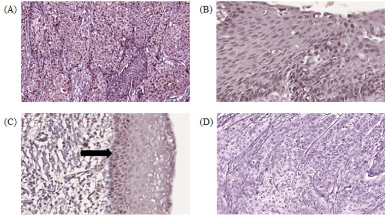
Figure 2.
Digital Scanned Images of MCM2 Immunohistochemical Staining Showing Brown Nuclear Staining in Cervical Epithelial Cells. (A) squamous cell carcinoma (SCC) showing high histoscore, (B) high-grade squamous intraepithelial lesion (HSIL) with high histoscore, (C) Normal cervix with low histoscore, positive staining in basal cells (arrows), (D) squamous cell carcinoma (SCC) with negative expression (objective x 20)

