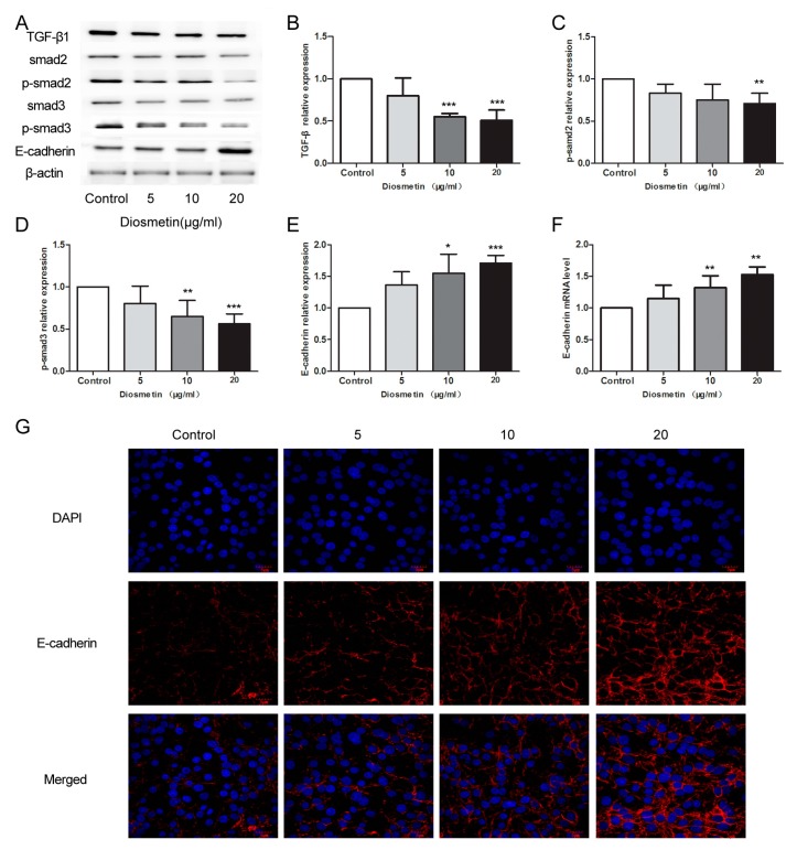
Figure 4.
Diosmetin led to inhibition of the TGF-β signaling pathway and activation of E-cadherin expression in glioma cells. Western blot assay was performed to detect the protein levels of (A,B) TGF-β1, (A,C) p-smad2, (A,D) p-smad3 and (A,E) E-cadherin, with the grayscale analysis using β-actin as the internal control. (F) Real-time PCR was used to detect the expression level of E-cadherin mRNA. (G) Immunofluorescence staining was performed to observe the distribution of E-cadherin protein in glioma cells. Compared with control group, * p < 0.05, ** p < 0.01, *** p < 0.001.

