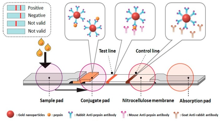
Figure 1.
The schematic diagram for the detection process of the pepsin immunochromatographic strip. To start a test, we apply a sample containing the analyte (i.e., pepsin) to the sample pad, and it subsequently migrates to the other parts of the strip. At the conjugate pad, pepsin is captured by gold nanoparticle (AuNP)-antibody conjugate. This pepsin-binding conjugate reaches the nitrocellulose membrane and moves under capillary action. At the test line, the pepsin-binding conjugate is captured by another antibody (monoclonal) that is primary to pepsin. Excess AuNP-antibody conjugate will be captured at the control line by secondary antibody. The colorimetric intensity at the test line, which corresponds to the amount of pepsin in saliva samples, is captured using a digital camera and analyzed using ImageJ software. The appearance of color at the control line ensures that a strip is functioning correctly.

