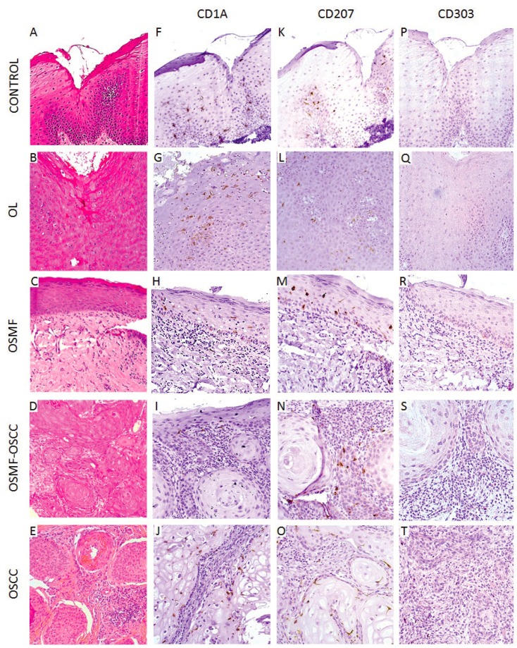
Figure 2.
Histopathological features and immunohistochemical detection of immature dendritic cells (DCs) and Langerhans cells (LCs) of control group (mucocele), oral leukoplakia (OL), oral submucous fibrosis (OSMF), OSMF associated with oral squamous cell carcinoma (OSMF-OSCC) and oral squamous cell carcinoma (OSCC). (A) Histologically normal oral epithelium without dysplastic alterations (hematoxylin and eosin – H&E; 200x). (B) Microscopic features of OL (H&E; 200x). (C) Microscopic features of OSMF demonstrating submucosal and justaepithelial deposition of collagenated connective tissue (H&E; 200x). (D) OSFM-OSCC with infiltrative features and cellular atipia (H&E; 200x). (E) OSCC demonstrating infiltrative tumor nests (H&E; 200x). (F) Immunohistochemical expression of CD1a+ cells in the epithelium tissue of the control group (mucocele). (G) OL group. (H) OSMF. (I) OSMF-OSCC. (J) OSCC. (K) CD207+ cells in the control group (mucocele). (L) OL group. (M) OSMF. (N)OSMF-OSCC. (O) OSCC. There was a remarkable decrease of CD1a+ and CD207+ cells in the OSCC group.

