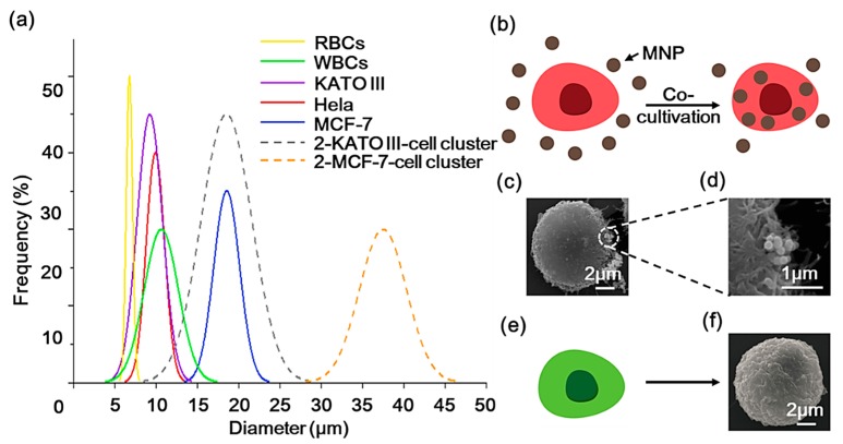
Figure 2.
(a) The diameters of some typical cells are summarized (solid lines). Dashed lines display the size distribution of the two types of circulating tumor cells (CTCs) (KATO III and MCF-7) clusters with two CTCs. (b) Illustrations of the co-cultivation process of the red fluorescent protein (Hela-RFP) cell with the magnetic nanoparticles (MNPs), and (c) the SEM image of the MNPs-labeled Hela-RFP. (d) SEM image of the surface attached of MNPs. (e) The green fluorescent protein (Hela-GFP) cells used to represent white blood cells (WBCs), and (f) the SEM image of the Hela-GFP cell morphology.

