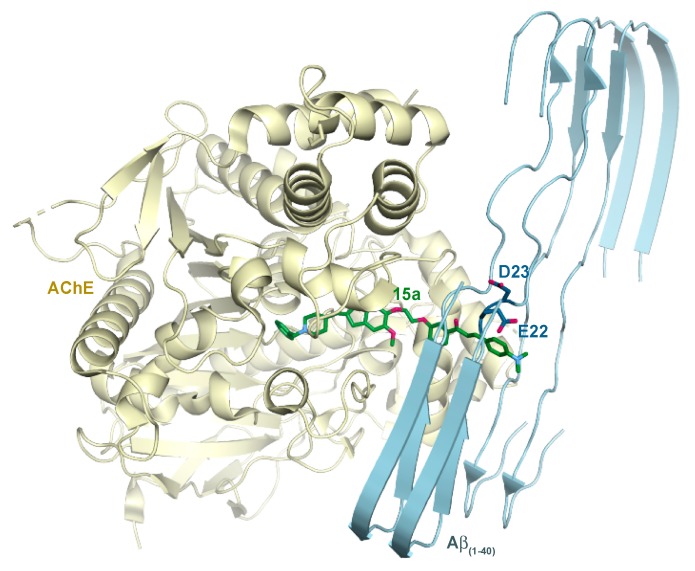
Figure 8.
A structural model of the HsAChE-15a-Aβ fibril complex. The HsAChE is shown in pale yellow, a two-layer Aβ (residues 1–40) fibril is in light blue, and 15a is in green. The acidic residues Glu22 and Asp23 of Aβ that may favorably interact with the dimethyl amino group of 15a are shown as dark blue sticks. The length of the linker of 15a can span no more than two β-strand layers, as shown in the figure. Note: Oxygen and nitrogen atoms are in red and blue, respectively.

