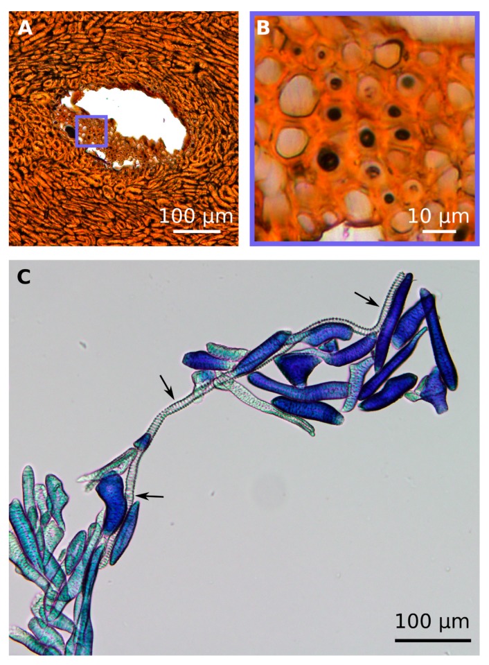
Figure 3.
LM images from the coconut endocarp showing tracheids. In the cross section of a vascular bundle ((A), polished thin section), the polygonal shape of tracheid cross-sections is visible (B). Macerated endocarp cells stained with Toluidin blue, showing an intact tracheid cell (⟶) with a length of 627 µm between accumulated sclereid cells (C).

