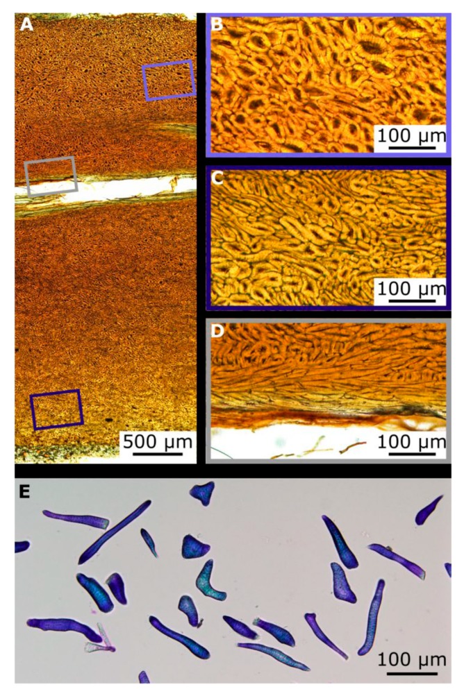
Figure 4.
LM images from the coconut endocarp showing sclereids. The cross section of a polished thin section shows a longitudinally cut vascular bundle within the brown tissue of the sclereid cells (A). Higher magnifications of the sclereid cell matrix reveal larger cells near the mesocarp side (B) than near the testa (C). Very long sclereids, the sclerenchyma fibers, are located in parallel to the vascular bundles (D). Macerated endocarp cells stained with Toluidin blue, showing sclereid cells with various shapes (E).

