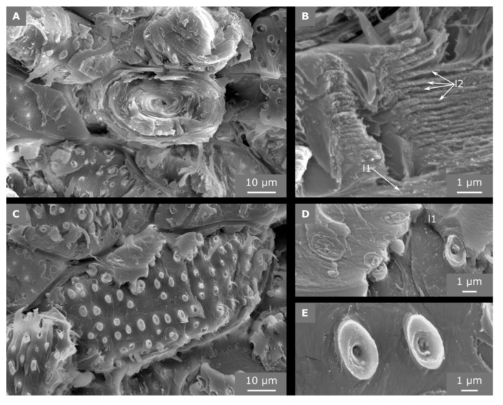
Figure 7.
SEM images of sclereid cells from fracture surfaces. The cross-section shows the many cell wall layers that fill almost the entire cell lumen (A). The cell wall is traversed by pit canals (B), to which the lignified secondary cell wall layers (l2) align. In a sclereid cell oriented longitudinally to the direction of fracture, the crack has run into the cell wall and detached the outermost cell wall layers (C). When the primary cell wall (l1) and middle lamella is partly broken, the pit cavity becomes visible (D) and the bordered pits emerge from the fracture surface (E).

