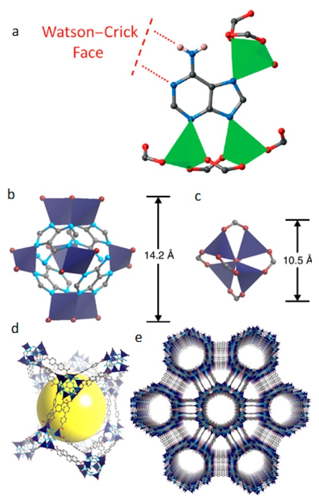
Figure 3.
(a) Open Watson–Crick sites and the coordination environment of adenine in ZnBTCA [59]. (b,c) A comparative illustration of the structure and size of the building units in bio-MOF-100 and the basic zinc-carboxylate building [57]. (d,e) The 3-D crystal structure of bio-MOF-100 where the cavities (yellow sphere) and the large channels can be seen (Zn2+: green or dark blue tetrahedra, C: grey spheres, O: red spheres, N: blue spheres, H: omitted for clarity) [57].

